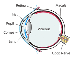
Typically when we go to an optometrist it is with the intention of improving eye sight. But, there is a lot more going on as we will learn. The frequently remembered portion of the eye exam is the test where we look at an eye chart and see how far down the chart that we can read. E, O, M, maybe Z, and in this test called refraction, the optometrist places lenses in front of each eye to determine the best corrective lens strength. This lens correction may be required when the light falling on the retina is either to far or too close due to the eye lens shape. The retina is the area inside of the eye that is like the photo element in a digital camera, or film in older cameras. It contains the rods and cones that allow the light signal to be sent to the optic nerve and provide vision. But, the retina also may contain clues to other problems unrelated to vision. For example, conditions like hypertension, diabetes, melanoma, detached retina and other problems may leave clues that allow the optometrist to suggest further testing leading to early diagnosis of an illness.
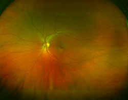
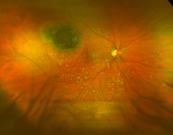
Four years ago I had eye surgery, and as a result I now have an annual examination with the surgeon. But, long before I signed up for the procedure I did the research to check the doctors credentials, background, and patient feedback. She is a well respected and patient supported professional so it was really a good choice to proceed with the necessary surgery. And, although I am using my own experience for this post, it is more about the technology and the importance of continuing eye care. Typically when we go to an optometrist it is with the intention of improving eye sight. But, there is a lot more going on as we will learn. The frequently remembered portion of the eye exam is the test where we look at an eye chart and see how far down the chart that we can read. E, O, M, maybe Z, and in this test called refraction, the optometrist places lenses in front of each eye to determine the best corrective lens strength. This lens correction may be required when the light falling on the retina is either to far or too close due to the eye lens shape. The retina is the area inside of the eye that is like the photo element in a digital camera, or film in older cameras. It contains the rods and cones that allow the light signal to be sent to the optic nerve and provide vision. But, the retina also may contain clues to other problems unrelated to vision. For example, conditions like hypertension, diabetes, melanoma, detached retina and other problems may leave clues that allow the optometrist to suggest further testing leading to early diagnosis of an illness. This photo is a scanning laser ophthalmoscopy image of a healthy retina from Optomap, a laser device from Optos Inc. Prior to the use of this technology the doctor would use an ophthlamoscope to examine the retina. But, the eye is usually dilated with a chemical to enlarge the pupil so that as much of the retina is exposed to examination as possible. With SLO, dilation is not necessary and the test is fast and painless. But, there are some caveats! This is the first time that I had an SLO done and it was with dilated eyes. So, while I was waiting for the doctor, the images of my eyes was displayed on a computer screen nearby. As I watched the image I noticed what the doctor had called a freckle or nevus. It was a clearly visible asymmetric spot among the capillaries and other features. It has not been a problem and continues to be more of an artifact. Also visible were the "floaters", those dark spots that we sometimes see move around in our visual field. Again, very normal. But, there is some controversy about needing SLO. This is an Optomap image of a retina showing melanoma as indicated by the small circular satellites in the lower center. These images are presumably fluorescent due to the dilating dye. The controversy about the use of SLO is that it only cover about 200 degrees in the typical 22 mm retina, which is about 72 % of the retinal area. This leaves the edges out of the image where disease can be found. So, I had an extensive conversation with the doctor about the potential use and need for SLO. Particularly important is that insurance may not cover the cost. My doctors take on the subject is that it is a valuable tool as a baseline metric but only as an adjunct to the opthalomoscope typically used for complete eye examination. So, for me, the takeaway is that we have to be well-informed patients and ask a lot of questions. Any good doctor expects questions and is prepared to explain any procedures. If they are not prepared, perhaps it may not be the right doctor for you. There is a link to this controversy here. Here's looking at you...
0 Comments
Leave a Reply. |
AuthorThe author has an eclectic background in chemistry, electronics, writing, mental health, and community action...Ken Archives
June 2021
|